Endocrine Disrupters and Genetically Modified Organisms Negate
Anti-Aging Protocols Leaving Only Specific Stem Cells as
Effective Counter Measures
Dr. Hans J. Kugler, PhD
Presented at the medical Congress of the American Academy of Anti-Aging Medicine, A4M, Orlando, FL, May 2012.
ABSTRACT
Longevity studies and scientific data clearly show that true life-long health, defined as a state of homeostasis according to Cheraskin/Kugler, can be achieved by correctly applying some 50 well-defined variables, which can also be grouped together as health practices. Recently published data regarding physical activity, the #1 essential and most effective requirement for true life-long health, and telomere research has made it possible to clearly and precisely define an anti-aging protocol. However, following such a precisely defined anti-aging protocol is negated by an ever increasing number of newly evolving obstacles, including endocrine disrupters and genetically modified organisms (GMOs). While some endocrine disrupters may be prevented or reversed with special detox programs, GMOs introduce risks that appear irreversible and only correctable with stem cells. Evaluating the various types of stem cells suggests that only individual-specific stem cells – nuclear transfer stem cells (NT-SC) – have the potential to be effective in treating/reversing GMO-caused damage, major diseases, spinal injuries, and more.
INTRODUCTION
In a series of 9 papers published 1974-86, Alabama University professor Emanuel Cheraskin, MD, DMD, PhD, defined true life-long health as a state of homeostasis, achieved by correctly applying some 50 variables, which can also be grouped together as health practices. Further cooperative research with Professor Cheraskin, performed in our lab, confirmed that life-long health is synonymous with optimum health, fitness, sexuality, best possible immune function, best recovery from disease or injury, and the lowest rate of cognitive decline and aging.1
Longevity Studies Confirm Cheraskin’s Definition of “Life-Long Health”
In longevity studies, performed at Roosevelt University in Chicago we divided cancer-prone mice – retired breeders – into 3 groups. Group 1: Nick-named “the average business man,” was given a junk food diet high in fat and sugar, with no supplements. This group did no exercise, and their environment was slightly stressed (small, standard cages), cigarette smoke was blown through the cages, and tap water given as drinking fluid. Group 2: The control group, everything was standard. Group 3: These mice were exercised in a rotating drum 3 times per week, were given a quality diet containing antioxidants, minerals, and supplements, drank carbon filtered water, and had larger living quarters with hiding places that were rearranged each time the cages were cleaned.
The result of the study showed a nearly 100% difference in the average lifespan between the animals in group 1 and 3, while group 2 was in-between. Professor Cheraskin had correctly predicted the outcome of our longevity studies. The results are in agreement with human longevity data. Synergism – as postulated by Cheraskin/Kugler – manifested itself in the longevity studies – and also in two “irreversible” heart recoveries – when several health practices were combined at the same time.2,3
The longevity studies, and Professor Cheraskin’s definition of health, were based on, and in agreement with, previously established theories of aging: glycation, free radical, cross-link, immunologic, and more. The theories of aging, and causes of premature aging, are discussed in the gerontology literature, and in an introduction to the theories of aging in chapter 11 of Life Long Health.4
Telomeres, Telomerase, and Anti-Aging
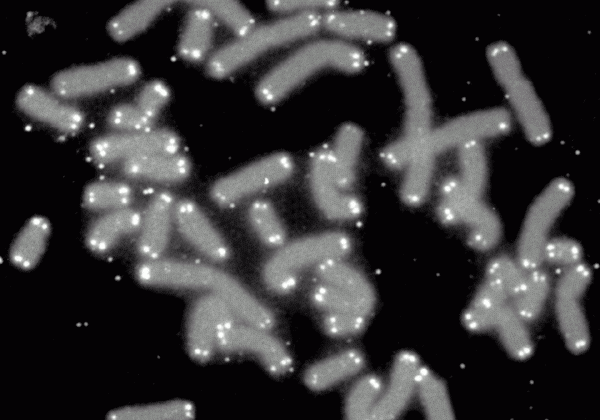
The limited lifespan theory of aging proposed by Professor Leonard Hayflick, (UC, San Francisco), was confirmed with the discovery of telomeres, protective elongated strands of repeating sub-units at the end of DNA/chromosomes. Telomeres are clear indicators of longevity. They are at their longest at birth, and they shorten by one sub-unit with each cell division. When a child is conceived, with both parents having shortened telomeres, the enzyme telomerase sets the telomeres in the fertilized egg back to full length.
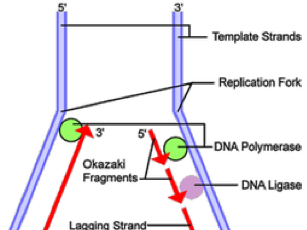
Interestingly enough, telomere science shows strong support for the intelligent design issue. Every species has telomeres; however different species have a different number of sub-units. Dogs have 15 (corresponding to a maximum lifespan of about 18 to 22 years), humans have 54 sub-units (maximum lifespan of 110-120), and the Galapagos tortoise has 75 (maximum lifespan 250 years). During cell division, enzymes that duplicate the chromosome and its DNA cannot continue their duplication all the way to the end of the chromosome (the so-called end replication problem). Therefore a sub-unit (see Okazaki Fragment in illustration below) is lost during each cell replication and telomeres get shorter.
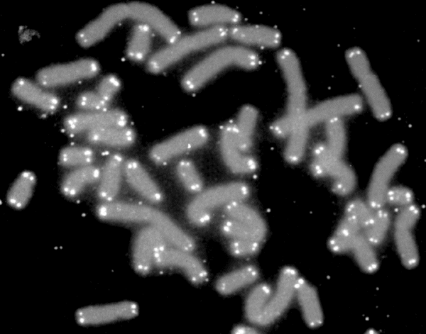
Figure 1. Human chromosomes (grey) are capped by telomeres (white).
Figure 2. Telomere shortening is a result of the end replication problem. DNA replication on the lagging strand results in the formation of Okazaki fragments.
Telomere length indicates remaining life potential. Therefore it is no surprise that scientists are exploring ways to lengthen aged telomeres. So far it has been established that, faulty health practices shorten telomeres (most likely due to affected telomerase activity), whereas good health practices lengthen them. Indeed, study results have shown that telomere lengthening can be achieved with exercise,5 good nutrition,6 supplements,7,8 and stress management.9
TA-65®
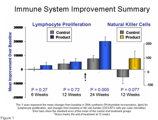
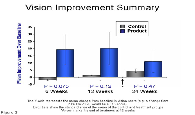
In the search for ways to increase telomere length, researchers from TA Sciences discovered that TA-65®, an extract from the Astralagus plant, showed impressive results never seen before, including lengthened telomeres, improvements in immune function, skin, vision and sexual functioning. Statistical evaluations were conducted at Stanford University. The results of these are reproduced in Figures 3 & 4.
Figure 3. TA-65® boosts immune function by increasing lymphocyte proliferation and the number of circulating natural killer cells.
Figure 4. TA-65® significantly improves vision score.
ENDOCRINE DISRUPTERS AND GENETICALLY MODIFIED ORGANISMS NEGATE THE EFFECTS OF AN ANTI-AGING PROTOCOL
It is clear that a scientifically documented and clearly defined path to anti-aging and optimum health has been established. Thus, following it should give the desired results. However, in many cases, from child development (premature puberty in boys and girls), to athletes (performance data), and regeneration in seniors, real results are often severely lacking or unobtainable. Why is this?
Research suggests that the potential benefits of following an anti-aging protocol are negated by environmental obstacles, such as endocrine disrupters and genetically modified organisms (GMOs). Indeed, presently available data suggests that many of the effects of endocrine disruptors and GMOs are severe and often irreversible.
The National Resources Defense Council (NRDC) defines an endocrine disruptor as a “synthetic chemical that when absorbed into the body either mimics or blocks hormones and disrupts the body’s normal functions. This disruption can happen through altering normal hormone levels, halting or stimulating the production of hormones, or changing the way hormones travel through the body, thus affecting the functions that these hormones control.” Recent research has shown that hormones play key roles in gene-expression – with as many as 20,000 receptors for estrogen alone – and that chemicals which mimic hormones interfere with, and block, normal gene expression, thus causing severe and extreme effects. Rachel Carson’s Silent Spring, pointing out the devastating effects of DDT,10 and Lindsey Berkson’s Hormone Deception,11 are just two books that called attention to medicine’s playing Russian roulette with untested chemicals.
GMOs (foods) introduce substances into our lives that, in their chemical actions, act like endocrine disrupters, block gene expression pathways, and even, often irreversibly, change life’s biochemical pathways. A lecture by Jeffrey Smith entitled Everything You Have to Know About Dangerous Genetically Modified Foods,12 provides a good introduction to the risks associated with GMOs.
As just one example of the risks associated with GMOs – demonstrating the severity of affecting/changing the most basic biochemical pathways – let’s take a detailed look at the two major types of genetically modified corn:
- Herbicide-resistant corn: A gene was introduced into the corn that makes it resistant against herbicide. The corn plants can now be sprayed with large amounts of herbicide (e.g. Roundup, atrazine) without affecting the corn. With this type of corn, you also ingest the herbicides. Atrazine, considered highly toxic, was banned in European countries years ago, while several studies have demonstrated the extremely toxic effects of Roundup. The herbicides, which are not quickly or easily degraded, also find their way into the environment where they severely poison ecosystems.
- Insect-resistant: A pesticide-producing gene was incorporated into the genetic structure of the corn. If you eat this type of corn, you also ingest lots of pesticide.
The list of medical disorders, and problems associated with the herbicides and pesticides – from digestive tract problems (human studies), to genetic aberrations (laboratory studies) – is literally growing on a daily basis.8,9 Recently, pesticides and herbicides have been found to cause hepatorenal toxicity,13 cytotoxicity in human cells,14 and cytotoxicity and DNA-damage in human-derived buccal epithelial cells.15 A large-scale study by Séralini et al found that rats fed a diet containing NK603 Roundup tolerant GM maize, or given water containing Roundup at levels permitted in drinking water and GM crops in the US, died earlier than rats fed on a standard diet. They suffered mammary tumors and severe liver and kidney damage.16
Both herbicides and pesticides are endocrine disrupters. Beyond that, research into the presence of GMO soy genes in the gut bacteria strongly suggests that genes from GMOs may wind up on our own genetic makeup.17,18 Sequences of genetically modified corn DNA have also been found in blood of cattle feeding on left-over corn stalks.19
Endocrine Disruptors at Work
Peptide synthesis (to trigger immune reactions, for example) offers a good example of how endocrine disruptors have negative effects on the body. Putting two-and-two-and-two of various research findings together, we can arrive with the below pathway for peptide synthesis:
- A messenger chemical – hormone, for example – arrives at the cell wall to transmit a message to the nucleus (DNA, inside the cell) that a certain peptide needs to be synthesized (Figure 5);
- Receptors are on the outside cell wall where the hormone fits in – if these receptors are blocked by an endocrine disrupter, the message can’t get passed along, or it starts a chain of reactions that are not required, resulting in endocrine disruption;
- If the message gets past along to the inside of the cell, a prostaglandin inside the cell picks up the message and transmits it to the nucleus. However, endocrine-disrupters can also interfere with prostaglandin synthesis, and if this happens, the message is interrupted again;
- If the message makes it to the DNA, based on the message carried by the prostaglandin, a gene on the DNA (that contains information about how to make the protein/peptide) is copied, resulting in an m-RNA (messenger RNA);
- The m-RNA travels to the r-RNA (ribosomal RNA, the workbench equivalent inside the cell) where the protein is assembled;
- t-RNAs (transfer-RNAs) – a different one for each amino acid – bring the amino acids to the workbench;
- The protein is assembled in the correct shape and fold – recall prions, the cause of mad cow disease, these are incorrectly folded proteins.
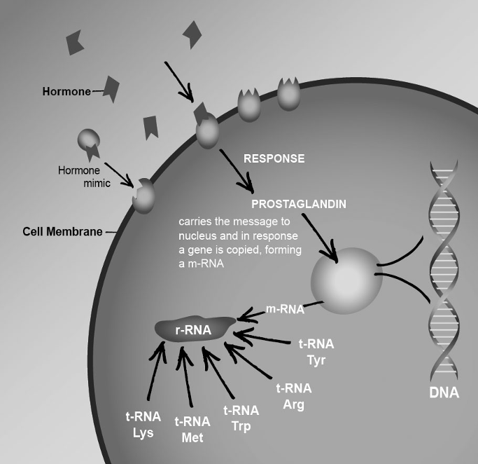
Figure 5. Multiple steps in peptide synthesis can be affected by endocrine disruptors.
The major response – mostly from supporters of fracking, environmental polluters, Monsanto and GMO stock holders (who else would support such Frankenfoods?) – used to be “well, you have some points there, but this (the lock and key receptors in the cell membrane, passing messages along to the inside in the cell, and then to the nucleus) is far from proven!”
The opponent’s arguments were silenced with the 2012 Nobel Prize for chemistry given to Drs. Robert Lefkowitz (Duke U) and Brian Kobilka (Stanford), showing that the pathway really is a “lock-and-key” mechanism; they call the receptors in the cell membrane that accept a message outside, and then pass it on in the inside as “G-protein-coupled receptors”, or GPCRs (it’s like a lock and key, Lefjkowitz said).
Problems, started by endocrine disrupters and/or GMOs, can be extremely severe, numerous, and irreversible. At present, two areas of research show promise:
- Detoxing: With increasing age, toxins that act as endocrine disrupters accumulate in adipose tissue; at age 40+, 50 to 100 endocrine disruptors, identifiable by name and chemical structure, have been identified in human adipose tissue. The niacin-based sauna detox can be effective in eliminating them;4
- Stem Cells: Eliminating the irreversible damage caused by GMOs appears – as of today’s state of the sciences – only possible with person-specific stem cells (NT-SC).20
STEM CELLS FOR TRUE ANTI-AGING AND REGENERATION
Presently available data suggest that only person-specific nuclear transfer (NT-SCs, with DNA the same as the recipient’s, and full telomere length) will be truly effective.
Adult Stem Cells
Research results with stem cells from various origins have demonstrated that adult stem cells are quite ineffective at regenerating organ functions via replacing aged or damaged tissues. This is most likely due to shortened telomeres that prevent further cell divisions. However, very impressive treatments – facial regeneration via sub-dermal implantation, regeneration of worn joints, heart recovery, prevention of limb amputation – have been reported at A4M Congresses by Dr. Sharon McQuillan, MD.21
Generic Embryonic Stem Cells
Generic embryonic stem cells (with DNA other than the recipient’s), harvested from aborted fetuses or from left-over embryos from fertility clinics) have been used in Europe on humans for several years now. Scientific publications and clinical feedback from European doctors have brought out two key findings:
- These stem cells give the body a noticeable boost, covering a wide spectrum from immune enhancement to general revitalization;
- However, these generic stem cells do not fully settle down in the recipient’s body, forming neither new tissues nor new organ cells.
Using stem cells (with DNA other than the recipient’s) is very much like an organ transplant; such cells have been shown to survive in a host body for a limited time period if immune-suppressants are used. I suggest that the overall positive results with generic stem cells are very similar to the results achieved with embryonic cell extracts, used in Germany.
Induced Pluripotent Stem Cells (iPS)
Induced pluripotent stem cells (iPS) are a type of stem cells artificially derived from a non-pluripotent cell, typically an adult somatic cell, by introducing control genes into the adult cell (skin, others) using retroviruses.22,23 This work in essence originated with the Japanese researcher Dr. Shinya Yamanaka who, together with British professor John B. Gurdon, won the 2012 Nobel Prize in Physiology. Interestingly enough, and in a departure from pure scientific facts, a key in giving this prestigious award to iPS was that – on the basis of religious arguments – occasional destruction of embryos could be avoided. Personally I believe that this concern is on most researchers minds; in my case I was raised in a catholic seminary school, and saving human embryos from destruction was the key in exploring the use of animal egg cells for preparing stem cells for therapeutic uses; nobody is cloning humans! iPS are of great scientific interest, but applications for human treatments are many years away for four major reasons:
- Risk of tumors, cancer24,25
- Telomere lengthening in iPS is questionable,
- Genetic abnormalities have been identified in iPS;26
- Rejection – iPS rejected by animal from which iPS were made.27
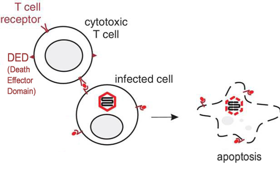
Would you agree to have such stem cells transplanted into your body? However, mechanism-wise, making cells with special characteristics – e.g. T-cells that can locate, and induce apoptosis in retrovirus-infected cells – this area looks very promising. For example, Rider et al developed a method called DRACO (double-stranded RNA (dsRNA) Activated Caspase Oligomerizer) to kill – very specifically – cells that are infected with viruses such as hepatitis-C, HIV, herpes, HPV, while leaving uninfected cells untouched.28 DRACO (Figure 6) works in the following way:
- The cytotoxic T-cell scours the body. When it finds dsRNA, it attaches to the cell;
- The death effector domain (DED) of the cytotoxix T cell then induces apoptosis (cellular suicide) in the infected cell;
- The cytotoxic T-cell moves on to find another infected cell.
Figure 6. Double-stranded RNA (dsRNA) Activated Caspase Oligomerizer (DRACO) selectively induces apoptosis in cells containing viral dsRNA, selectively killing infected cells without harming uninfected cells.
A fact of life: Stem cells, in order to be truly effective and settle down in the recipient’s body (and without any rejection responses), must have DNA the same as the recipient’s, and be highly active (full telomere length). The scientific basis for the need of NT-SCs was presented by this author at the 2012 Orlando A4M Congress. “Stem Cell Reality Check: The Truth and Myth”.29 The notion that reversal of spinal injuries will require NT-SCs has been supported by Swiss scientists at Lausanne University.30
Methods for Making Person-Specific Embryonic Stem Cells
Person-specific embryonic stem cells are achievable via 2 major methods:
- Nuclear Transfer; possibly with the use of animal oocytes for nuclear transfer;
- Parthenogenesis – triggering an oocyte to divide without fertilization, however this method is limited to women who had some of their egg cells collected and stored during their child-bearing years (stem cells will be specific only for the person from whom the egg cell originated).
On 15 March, 2008, in a news release, the International Academy of Alternative and Anti-Aging Medicine (IAAM) stated: “Parthenogenesis, Virgin-Birth: A way of fulfilling the stem cell needs of 50% of the population and without ethical objections. With much admiration for the ground-breaking work at Boston Children’s Hospital,31 Kobe University in Japan,32 SUMS University in China,33 the University of Milan, Italy,34 the University of South Florida, Tampa,35 Heinrich Heine University, Germany,36 Michigan State University,37 and many others:
We believe that, on the basis of available data, the time is right for IAAM Stem Cell Research, Dr. Hans J. Kugler, PhD, director, to recommend that woman of child-bearing age give it serious consideration to have a number of their egg cells collected and stored (by a cryogenic facility) for possible future need of stem cells.”
For men (and women who are not candidates for parthogenesis), making individual-specific stem cells involves the procedures performed at a nuclear transfer laboratory. How does a nuclear transfer work?
- The person who needs stem cells – because of a major disease, cancer, heart disease, Parkinson’s, Alzheimer’s, diabetes, or simply for anti-aging or rejuvenation procedures – supplies his DNA in the form of any cell, like a skin cell, to a NTL;
- The nucleus (DNA) is removed from the skin cell (or from any other cell), and is saved;
- A human egg cell is prepared for the nuclear (DNA) transfer by removing its nucleus;
- The previously saved patient’s nucleus (DNA) is transferred into the (denucleated) egg cell;
- The cell is triggered to start dividing. This is where we currently run into problems, as scientists have had lots of troubles with this important step. As of today only about one in 30 to 50 attempts succeed. Since human egg cells are also very expensive – $10,000 for 4 egg cells – this is quite a rate/cost-determining step. An electric shock method is used to trigger the cell to start dividing. With nature it is the sperm that enters the egg cell and then starts the process of cell divisions, lots of interesting chemistry, but no electric shocks. At HK Stem Cell Research we believe that there are more effective methods for stimulating mitosis, and some initial approaches have already been shown effective;
- After cell division – mitosis – is initiated, on day 4, the blastocyst stage, stem cells start forming in a small sac in the blastocyst. These stem cells have the potential to further develop into any kind of cells, e.g. heart, muscle, skin, kidney, liver, brain, whole organs;
- The stem cells are removed from the blastocyst and are separately cultivated in a special CO2 incubator;
- The stem cells are administered to the patient; the procedure of doing so is very simple. The implanted stem cells have a way of sensing where repair is needed, and will travel there, carried in the bloodstream. The cells can also be injected into a specific organ.
Besides the many negative findings about iPS, there is some encouraging stem cell research in favor of nuclear transfer procedures. Byrne et al demonstrated on Rhesus monkeys that a nuclear transfer – using DNA from a skin cell – is possible and does yield stem cells.38 Data clearly demonstrate that embryonic stem cells already hold the key to curing Parkinson’s disease.39-41 By following the nuclear transfer approach to make specific embryonic stem cells it is hoped that these findings will be replicated in humans.
Because human egg cells are very expensive, scientists have considered using animal egg cells. The use of animal egg cells, like bovine or rabbit egg cells (oocytes), is a definite possibility; after all, with the DNA removed, the rest is just like fertile soil. Think of it this way: You can grow corn on crushed Hawaiian volcanic rock, on beach sand, or on any type of soil, as long as water and nutrients are supplied; the result will always be corn. This possibility has recently been evaluated and published in a major paper, authored together by British and US research groups.42 Embryonic stem cells have also been successfully made via nuclear transfer of human DNA into rabbit oocytes.43 This could possibly be of special interest for spinal cord injuries.
Embryonic Cell Extracts
During a collaborative 8-year study (1985-93) Tuebingen and Heidelberg universities in Germany, evaluated and refined a treatment known as “Dr. Niehans Zelltherapie”, during which patients were given organ-specific cells from embryonic sheep tissues, and pinpointed organ-specific low molecular weight nucleic acids, peptides and growth factors as the active ingredients (now used in Germany as injectable organ-specific embryonic “cell extracts” in the treatment, or adjunct, in many diseases). More basic forms of such low molecular weight nucleic acids, peptides and growth factors can also be found in the generic stem cells; facts confirmed during a visit (2005) to Germany, when I visited with Dr.Ulrich Friedrichson, MD, PhD, one of the lead researchers in the above mentioned studies. These cell extracts (heart, muscle and mesenchyme) were also used in my own full recovery from a heart injury for which, cardiologists had advised, treatment was impossible. The heart recovery protocol was presented at the 17th Medical Congress of A4M in Las Vegas, December 2009,2 and confirmed with the recovery of another male patient who was on an emergency heart transplant list, the full details of this heart recovery protocol are included in the book Anti-Aging Medical Therapeutics Volume XII.3 Latest treatment protocols, mechanism of action, and safety of the cell extracts were presented by Dr. Friedrichson, MD, PhD, at the Bangkok Anti-Aging Congress, September 2011.44
If we had person-specific embryonic stem cells (psESCs, DNA same as recipient’s) today, and applied them via a number of pathways, the stem cells would regenerate organs and cause an enhanced production of organ-specific low molecular weight nucleic acids, peptides, and growth factors, thus resulting in functioning of the organs at near-perfect. Since we don’t have psESCs yet, injecting organ-specific cell extracts is the present most promising way of enhancing organ function.
Back to Basics; Checking Your Anti-Aging Status
No matter at what point we are – perfectly applied anti-aging principles, toxic substances eliminated with detox, or tissues regenerated with stem cells – a follow-up to check if we are truly on the right track should be part of anybody’s anti-aging program. How can this be done?
In the future the Bio-Chip, measuring gene expression during a doctor visit, will, most likely, be a key tool in evaluating a person’s health status.4B Presently there is a fast, reliable and non-invasive 3D Full Body Assessment System available to monitor aging processes of the body. As discussed at A4M meetings by Dr. Luba Diangar, it is non-invasive and doesn’t use radiation or ultrasound.45
CONCLUSIONS
Anti-aging protocols – supported by longevity studies on animals and humans, confirmed with disease statistics, and in agreement with the theories on aging – have been evolving to a point where most aspects of “how to” anti-aging programs are precisely defined. Statistics even allow us to calculate the percent-effectiveness of anti-aging protocols for people who are incapable or unwilling, to abide by essential anti-aging requirements, like unable to exercise, remain overweight, insist on smoking, or are unwilling to give up a practice that has clearly established negative effects on health and anti-aging.
With the newly evolving obstacles – endocrine disrupters and GMOs – the scientific data are clear: they represent great risks to human health, and negate anti-aging and life-long health achievements. That such harmful substances find their way into our environment and foods is clearly due to special interest crimes/abuse: The crimes continue, only the names of the criminals have changed: instead of DDT and DES, we now have BPA, phthalates, GMOs, fracking chemicals, and toxic metals. Instead of researching new anti-aging protocols that include these new obstacles, we must follow a logical approach, and place a heavy emphasis on ways to pinpoint such problem areas at the earliest stage possible, and find ways to eliminate them from our lives. At the present time, key priorities would be to prevent poisoning the environment with toxic chemicals, eliminate fracking chemicals, establish protocols to prevent dangerous GMOs from getting into the food supply, and make it a requirement not to assume safety, but to follow safety protocols to establish safety for any new modality that is to be introduced into our environment or human/animal health.
ABOUT THE AUTHOR
Dr. Hans J. Kugler, PhD, is President and founder of the International Academy of Anti-Aging Medicine and HK Stem Cell Research, and past president and board member of the National Health Federation. He received his “Vordiplom” (BS equivalent) in physiology at the University of Munich Medical School under Nobel Laureate A. Butenandt, PhD in chemistry SUNY, Stony Brook, NY, was assistant professor at Stony Brook, and associate professor at Roosevelt University, Chicago, teaching pre-meds and doing aging research with cancer-prone animals. Residing in Redondo Beach, southern California, his present research focuses on NT stem cells, and natural apoptosis complexes. As author of 8 books on anti-aging, health and fitness – most recent e-book “LIFE-LONG HEALTH: Learn how to Control your Genes to stay Young with Age” – and having made medical history with his own heart recovery protocol – presented at A4M’S 2009 Congress – Dr. Kugler does anti-aging counseling at the Health Integration Center in Torrance, California, is a frequent presenter at anti-aging medical meetings, and is available as motivational speaker about HOW TO anti-aging from the ground up; from fitness, to weight-loss, environment, detoxing, alternative medicine, and key anti-aging modalities.
Contact: drkugler1@verizon.net or visit www.antiagingforme.org
REFERENCES
- Kugler H. The Scientific basis for defining pure life-long health as a state of homeostasis, with telomere, stem cell and aging research confirming health practices as true anti-aging modalities. Paper presented at: 17th Medical Congress of the American Academy of Anti-Aging Medicine; April 7-9, 2011; Orlando, FL.
- Kugler H. Alternative treatments in cardiac injury. Paper presented at: 17th Medical Congress of the American Academy of Anti-Aging Medicine; December 11, 2009; Las Vegas, NV. CD available at: http://www.instatapes.com/A4M/toc.htm , Disc 3, Talk No.2.
- Kugler H. Friedrichson U, Ghaly F, Ward P. Alternative therapies in the treatment of cardiac injury; a case report with recovery regimens for non-ablatable atrial fibrillation and greatly enlarged left atrium. In: Klatz R, Goldman R, eds. Anti-Aging Medical Therapeutic, Volume XII; 2010:173-180.
- A) Kugler H. The Scientific Basis for Developing a Personal Anti-Aging Program. In: Klatz R, Goldman R, eds. Anti-Aging Medical Therapeutics Volume. IV. A4M. B) Kugler H. Life-Long Health: Learn how to Control you Genes to Stay Young with Age. Available at: http://www.antiagingforme.com .
- LaRocca TJ, Seals DR, Pierce GL. Leukocyte telomere length is preserved with aging in endurance exercise-trained adults and related to maximal aerobic capacity. Mech Ageing Dev. 2010;131:165-167.
- Ornish D, Lin J, Daubenmier J, et al. Increased telomerase activity and comprehensive lifestyle changes: a pilot study. Lancet Oncol. 2008;9:1048-1057.
- Xu Q, Parks CG, DeRoo LA, Cawthon RM, Sandler DP, Chen H. Multivitamin use and telomere length in women. Am J Clin Nutr. 2009;89:1857-1863.
- Houston D. Vitamin D and physical function in older men and women. Paper presented at: 2010 Experimental Biology Meeting; April 25, 2010.
- Epel ES, Blackburn EH, Lin J, Dhabhar FS, Adler NE, Morrow JD, Cawthon RM. Accelerated telomere shortening in response to life stress. Proc Natl Acad Sci U S A. 2004;101:17312-17315.
- Carson R. Silent Spring. Houghton Miflin; 1962.
- Berksen DL. Hormon Deception. McGraw-Hill Prof Med/Tech; 2001.
- Smith J. Everything You Have to Know About Dangerous Genetically Modified Foods. Available at: http://vimeo.com/6575475. Last accessed October 4, 2012.
- Séralini GE, Cellier D, de Vendomois JS. New analysis of a rat feeding study with a genetically modified maize reveals signs of hepatorenal toxicity. Arch Environ Contam Toxicol. 2007;52:596-602.
- Mesnage R, Clair E, Gress S, Then C, Székács A, Séralini GE.Cytotoxicity on human cells of Cry1Ab and Cry1Ac Bt insecticidal toxins alone or with a glyphosate-based herbicide. J Appl Toxicol. 2012 Feb 15. doi: 10.1002/jat.2712.
- Koller VJ, Fürhacker M, Nersesyan A, Mišík M, Eisenbauer M, Knasmueller S. Cytotoxic and DNA-damaging properties of glyphosate and Roundup in human-derived buccal epithelial cells. Arch Toxicol. 2012;86:805-313.
- Séralini GE, Clair E, Mesnage R, Gress S, Defarge N, Malatesta M, Hennequin D, de Vendômois JS. Long term toxicity of a Roundup herbicide and a Roundup-tolerant genetically modified maize. Food Chem Toxicol. 2012 Sep 11. pii: S0278-6915(12)00563-7. doi: 10.1016/j.fct.2012.08.005.
- Netherwood T, Martín-Orúe SM, O’Donnell AG, Gockling S, Graham J, Mathers JC, Gilbert HJ. Assessing the survival of transgenic plant DNA in the human gastrointestinal tract. Nat Biotechnol. 2004;22:204-209.
- Ferrini AM, Mannoni V, Pontieri E, Pourshaban M. Longer resistance of some DNA traits from BT176 maize to gastric juice from gastrointestinal affected patients. Int J Immunopathol Pharmacol. 2007;20:111-118.
- Bertheau Y, Helbling JC, Fortabat MN, et al. Persistence of plant DNA sequences in the blood of dairy cows fed with genetically modified (Bt176) and conventional corn silage. J Agric Food Chem. 2009;57:509-516.
- Kugler H. Stem cells facts and fiction: What needs to be done? Paper presented at: A4M Anti-Aging Congress; May 17, 2012; Orlando, FL.
- McQuillan S. Clinical outcomes of stem cell therapy for myocardial ischemia and congestive heart failure. Paper presented at: A4M Anti-Aging Congress; May, 2011; Orland. FL.
- Takahashi K, Yamanaka S. Induction of pluripotent stem cells from mouse embryonic and adult fibroblast cultures by defined factors. 2006;126:663-676.
- Okita K, Ichisaka T, Yamanaka S. Generation of germline-competent induced pluripotent stem cells. 2007;448:313-317.
- Fujioka T, Shimizu N, Yoshino K, Miyoshi H, Nakamura Y. Establishment of induced pluripotent stem cells from human neonatal tissues. Hum Cell. 2010;23:113-118. doi: 10.1111/j.1749-0774.2010.00091.x
- Ohm JE, Mali P, Van Neste L, et al. Cancer-related epigenome changes associated with reprogramming to induced pluripotent stem cells. Cancer Res. 2010;70:7662-7673.
- Hayden EC. Stem cells: The growing pains of pluripotency. 2011;473:272-274. Available at: http://www.nature.com/news/2011/110518/full/473272a.html. Last accessed October 4, 2012.
- Zhao T, Zhang ZN, Rong Z, Xu Y. Immunogenicity of induced pluripotent stem cells. 2011;474:212-215. doi: 10.1038/nature10135.
- Rider TH, Zook CE, Boettcher TL, Wick ST, Pancoast JS, Zusman BD. Broad-spectrum antiviral therapeutics. PLoS One. 2011;6:e22572.
- Kugler H. Stem cells facts and fiction: What needs to be done? Paper presented at: A4M Anti-Aging Congress; May 17, 2012; Orlando, FL.
- Dominici N, Keller U, Vallery H, et al. Versatile robotic interface to evaluate, enable and train locomotion and balance after neuromotor disorders. Nat Med. 2012;18:1142-1147.
- Lengerke C, Kim K, Lerou P, Daley GQ. Differentiation potential of histocompatible parthenogenetic embryonic stem cells. Ann N Y Acad Sci. 2007;1106:209-218.
- Ogushi S, Palmieri C, Fulka H, Saitou M, Miyano T, Fulka J Jr. The maternal nucleolus is essential for early embryonic development in mammals. 2008;319:613-616.
- Mai Q, Yu Y, Li T, Wang L, Chen MJ, Huang SZ, Zhou C, Zhou Q. Derivation of human embryonic stem cell lines from parthenogenetic blastocysts. Cell Res. 2007;17:1008-1019.
- Brevini TA, Gandolfi F. Parthenotes as a source of embryonic stem cells. Cell Prolif. 2008;41 Suppl 1:20-30.
- Liu L, Bailey SM, Okuka M, et al. Telomere lengthening early in development. Nat Cell Biol. 2007;9:1436-1441.
- Fangerau H. Can artificial parthenogenesis sidestep ethical pitfalls in human therapeutic cloning? An historical perspective. J Med Ethics. 2005;31:733-735.
- Cibelli JB, Cunniff K, Vrana KE. Embryonic stem cells from parthenotes. Methods Enzymol. 2006;418:117-135.
- Byrne JA, Pedersen DA, Clepper LL, Nelson M, Sanger WG, Gokhale S, Wolf DP, Mitalipov SM. Producing primate embryonic stem cells by somatic cell nuclear transfer. 2007;450:497-502.
- Kim JH, Auerbach JM, Rodríguez-Gómez JA, et al. Dopamine neurons derived from embryonic stem cells function in an animal model of Parkinson’s disease. 2002;418:50-56.
- Fathi F, Altiraihi T, Mowla SJ, Movahedin M. Transplantation of retinoic acid treated murine embryonic stem cells & behavioural deficit in Parkinsonian rats. Indian J Med Res. 2010;131:536-544.
- Amano T, Papanikolaou T, Sung LY, Lennington J, Conover J, Yang X. Nuclear transfer embryonic stem cells provide an in vitro culture model for Parkinson’s disease. Cloning Stem Cells. 2009;11:77-88.
- Chung Y, Bishop CE, Treff NR, et al. Reprogramming of human somatic cells using human and animal oocytes. Cloning Stem Cells. 2009;11:213-223.
- Chen Y, He ZX, Liu A, et al. Embryonic stem cells generated by nuclear transfer of human somatic nuclei into rabbit oocytes. Cell Res. 2003;13:251-263.
- Friedrichson U. Treatment protocols, and mechanism of actions, with embryonic cell extracts. Paper presented at: Anti-Aging Congress; May, 2011; Bangkok, Thailand. Contact: info@friedrichson.de .
- Diangar Luba, DNM, LD Diagnostics, Costa Mesa, CA, 2Steps2Health.com .

Leave a Reply